Case Reports - Journal of Basic and Clinical Reproductive Sciences (2017) Volume 6, Issue 2
Successful Myomectomies during Pregnancy
Received: 10-May-2017 Accepted Date: Dec 04, 2017 ; Published: 07-Dec-2017
Citation: Chakraborty S, Maji S and Ray I.Successful Myomectomies during Pregnancy doi: 10.4103/2278-960X.194510
This open-access article is distributed under the terms of the Creative Commons Attribution Non-Commercial License (CC BY-NC) (http://creativecommons.org/licenses/by-nc/4.0/), which permits reuse, distribution and reproduction of the article, provided that the original work is properly cited and the reuse is restricted to noncommercial purposes. For commercial reuse, contact reprints@pulsus.com
Introduction
The prevalence of leiomyoma during pregnancy is reported as 2% [1]. During pregnancy, uterine leiomyoma are usually asymptomatic but may be occasionally complicated by red degeneration and an increased frequency of spontaneous abortion, preterm labour, premature rupture of fetal membranes, antepartum haemorrhage, malpresentations, obstructed labour, caesarean section and postpartum haemorrhage [1-3].
The management of uterine leiomyoma during pregnancy is largely expectant and its surgical removal is generally delayed until after delivery [4-7]. Because of the increased vascularisation of the uterus during pregnancy, women are at increased risk of bleeding and postoperative morbidity during myomectomy [2,5,6,8,9]. Some reports have shown that myomectomy during caesarean delivery can be safe [7,10-15]. Controversy persists among reports of myomectomy being performed during pregnancy [1], with some case series having reported the safety of antepartum myomectomy in carefully selected patients [17-20].
We present 3 cases of large symptomatic fibroid diagnosed during pregnancy which was successfully managed by antepartum myomectomy.
Case Presentation
History, examination and management
A 34-year old primigravida presented to our hospital on 6th November 2014 with 3 month history of abdominal swelling and amenorrhea of 13 weeks duration. The abdominal swelling started as a small lump but markedly increased in size in the preceding 2 months. It was associated with pain, severe epigastria discomfort and constipation.
The patient was mild pale and had bilateral pitting pedal edema. The pulse rate was 80 beats per minute and the blood pressure was 120/70mmHg. The respiratory rate was 24 cycles per minute. The abdomen was grossly distended and tense and reaching up to epigastric region. There was a massive central Abdomino-pelvic mass which was firm and irregular, measuring 40 cm from the symphysis pubis.
Abdominal sonography showed an intra-uterine viable singleton fetus of 13 weeks 6 days gestation and a huge fundal fibroid 22 × 19 cm extending up to epigastrium.
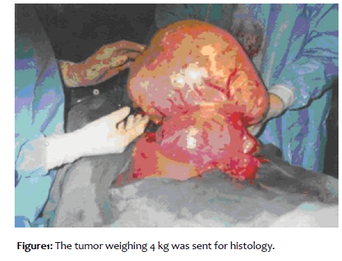
Blood tests showed a Hb 9.4 gm% and normal electrolytes, urea and creatinin levels. The woman's blood group was A Rhesus positive decision of myomectomy taken because of the severity of symptoms. Laparotomy was performed under general anaesthesia with endotracheal intubation on 18th nov 2014.the peritoneal cavity was entered through mid-line vertical incision. A large myoma identified at fundus of uterus of size 20 × 20 cm. A transverse incision given at fundus and base of myoma and myoma is removed by sharp and blunt dissection and with help of myoma screw. Endometrial cavity is not entered and uterus is closed with vicryl 2-0 with baseball suture. A huge myoma of size 20 × 20 cm weighing 4 kg is sent for histopathology. The uterus was soft and compatible with 16 weeks pregnancy. The ovary and tubes were grossly normal. The post operating event was uneventful and the patient received 4 units of packed red blood cells. A post-operative ultrasonography was repeated on 14th post-operative day and revealed a single live fetus of approximately 21 weeks 2 days gestation. Patient was discharged on 14th post-operative day and advised for antenatal follow up and HPE report her HPE report initially shows Leiomyosarcoma because of Nuclear pleomorphic and hypercellularity.It was reviewed again in another centre whose HPE report shows Leiomyoma as the above features can be normal in pregnancy [Figure 1].
Intramuscular ritodrine given 2 doses one just before operative day and one on 1st post-operative day along with weekly intramuscular proluton to prevent uterine contractions and the woman had an uneventful postoperative follow up. She was booked for antenatal care and had regular follow up. She underwent elective caesarean at 38 weeks 1 day gestation with delivery of a live healthy female baby weighing 2.75 kg. The puerperium was uneventful. The 6 weeks post-natal visit was unremarkable.
Case 2
A 32 yrs old primigravida admitted throughout patient department with complain of progressive abdominal swelling along with 18 weeks of Amenorrhoea on 23rd may 2015. She was having abdominal distension which was irregular in shape and corresponding to 36 weeks size of uterus. her ultrasonography reveals a fibroid in fundal region towards left side measuring 17.1 × 11.1 × 15.3 cm along with an intrauterine pregnancy of 16 weeks 4 days gestation. She underwent myomectomy on 26th may 2015.laparotomy was done with mid line vertical incision. On opening peritoneum multiple myoma seen a large myoma of size 18 × 12cm seen in posterior wall and a small Myoma of size 4 × 3 cm seen. Posterior wall myoma is removed with myoma screw with blunt and sharp dissection. Uterine cavity not entered. Uterine myometrium closed with vicryl 1 and serosa is closed with baseball suture with vicryl 2-0. Uterus size corresponding with 16 weeks size. Bilateral tubes and ovary healthy. Haemostasis secured. Specimen weighing 2 kg is sent for histopathological examination. Injection ritodrine was given 1 day before operation and on 1st post-operative day intramuscularly and injection proluton 250 mg weekly in dose. Post-operative event was uneventful and she received one unit of packed red cell. Her ultrasonography was done on 7th post-operative day which revealed single live fetus with apparent gestational age of 18 weeks 3 days. She was discharged on 10th post-operative and advised to follow up for routine antenatal checkup. Her HPE report show leiomyoma. She underwent elective caesarean at 37 weeks gestation with delivery of a healthy male baby of weight 3 kg. Her puerperal period was uneventful.
Case 3
A 23 years old primigravida admitted through emergency with severe abdominal pain on 14June, 2015 with 15 weeks of Amenorrhoea. she was ill looking mild pale with abdominal distension, irregular margin corresponding to 30 weeks gestation. She was managed conservatively with analgesics for pain abdomen. Her ultrasonography reveals single live fetus with 14 weeks 1 day gestation and a large myoma in the anterior wall of uterus of six 10x8 cm. her pain subsided with analgesics. She was conservatively managed for 1 month. Her ultrasonography was repeated again which show an intrauterine live fetus of 17 weeks gestation and multiple fibroids with one large fibroid on anterior wall compressing the gestational sac. She was planned for myomectomy on 14july, 2015 under GA. Abdomen was opened with vertical incision on opening peritoneum a large myoma seen on anterior wall of uterus of size 12 × 10 cm and another on right lateral wall of size 8 × 6 cm. both myoma was removed with sharp and blunt dissection using myoma screw. uterine cavity not opened. Uterine myometrium closed and serosa closed with baseball sutures. Uterus was of size of 18 weeks. Abdomen closed in layers. Both the myomas weighing approx 2 kg was sent for histopathological examination. Patient was given injection ritodrine on post-operative day along with injection proluton weekly intramuscularly. Post-operative period was uneventful. She was discharged on 10th postoperative day and advised for routine antenatal check-up. Her HPE report shows leiomyoma. She was electively admitted at 36 weeks of gestation. Her elective caesarean section was performed at 36 weeks 2 days gestation after giving intramuscular betamethasone 24 hours prior to operation. She delivered a healthy boy baby weighing 2.3 kg. Her puerperal period was uneventful.
REFERENCES
- Lolis DF, Kalantaridou SN, Makrydimas G, Sotiriadis A, Navrozoglu I et al (2015) Successful myomectomy during pregnancy. Human Reproduction.18:1699–1702.
- Brown D, Fletcher HM, Myrie MO, Reid M.(1999) Caesarean myomectomy- a safe procedure. A retrospective case controlled study. Obstet Gynecol.19:139–141.
- Ikedife D.(1999) Surgical challenge of myomectomy at cesarean section. Nigerian Journal of Surgical Sciences. 3:15–17.
- Kwawukume EY. (2012) Myomectomy during caesarean section. Int J Gynecol Obstet.
- 76:183–184.
- Ezechi OC, Kalu BKE, Okeke PE, Nwokoro CO. Inevitable caesarean myomectomy: A case report. Trop J Obstet Gynecol. 20:159–160.
- Ehigiegba AE, Evbuomwan CE.(1998) Inevitable caesarean myomectomy. Trop J Obstet Gynecol.15:62.
- Roman AS, Tabsh KM.(2014) Myomectomy at time of cesarean delivery; a retrospective cohort study. BMC Pregnancy and childbirth. 4:14.
- Depp R. Caesarean delivery. In: Gabbe SG, Niebyl JR, Simpson JL, editor. Obstetrics: normal and problem pregnancies. 4. New York: Churchill Livingstone 599.
- Cunningham FG, Gant FN, Levenok KJ, Gilstrap LC, Hauth JC et al (2000) editors . Williams Obstetrics. 21. New York: McGraw Hill Chapter35: Abnormalities of the reproductive tract; pgno. 930.
- Michalas SP, Oreopoulou FV, Papageorgiou JS (1995) Myomectomy during pregnancy and caesarean section. Human Reproduction.10:1869–70.
- Sapmaz E, Celik H, Altungul (2003) A Bilateral ascending uterine artery ligation vs. tourniquet use for haemostasis in caesarean myomectomy. A comparison. J Reprod Med.48:950–954.
- Ehigiegba AE, Ande AB, Ojobo SI. (2001) Myomectomy during cesarean section. Int J Gynecol Obstet.75:21–25. doi: 10.1016/S0020-7292(01)00452-0.
- Cobellis L, Pecori E, Cobellis G.(2002) Hemostatic technique for myomectomy during caesarean section. Int J Gynecol Obstet. 79:261–262. doi: 10.1016/S0020-7292(02)00299-0.
- Ortac F, Gungor M, Sonmezer M(1999) Myomectomy during caesarean section. Int J Gynecol Obstet. 67:189–190. doi: 10.1016/S0020-7292(99)00129-0.
- Kwawukume EY. (2002) Caesarean myomectomy. Afr J Reprod Health. 6:38–43.
- Burton CA, Grimes DA, March CM (1998) Surgical management of leiomyomata during pregnancy. Obstet Gynecol. 74:707.
- Joo JG; Inovay J, Silhavy M, Papp Z (2001) Successful enucleation of a necrotizing fibroid causing oligohydramnios and fetal postural deformity in the 25th week of gestation. A case report. J Reprod Med 46:923–925.
- Low SC, Chong CL.(2004) A case of cystic leiomyoma mimicking an ovarian malignancy. Acad Med Singapore. 33:371–374.
- Exacoustos C, Rosati P (1993) Ultrasound diagnosis of uterine myomas and complications in pregnancy. Obstet Gynecol 82:97–101.


